Radiation Oncology (Physics)
Basic Therapy Physics
Radiation physics
Treatment Planning
Basic treatment planning
3-D conformal therapy
3-D conformal & IMRT
Imaging in treatment planning
Intensity modulated RT (IMRT)
Inverse treatment planning
CT-simulation
Imaging
Basic principle
3D
CT
MRI
PET
Ultrasound
IGRT
Lung
Neurosurgery
Prostate
Dosimetry
General
Intraoperative
Photon & electron
Monitor Unit
Neutron
Brachytherapy
General
Intra-vascular
High-dose rate
Low-dose rate
Monte carlo dosimetry
Pediatric
Quality assurance
Dosimetrist
Physics
Clinical
Brachytherapy
CT-Simulation
Radiobiology
Radiation Therapists
Physics
Clinical
Radiation protection
Special Procedures
Intraoperative
Hyperthermia
Neutron capture therapy
Stereotactic radiosurgery
Linear Accelerator
Radiation & Cancer Biology Practice Examination
Radiation & Cancer Biology Practice Examination
Rabex 2025
Basic
Rabex 2025
Applied
Clinical
Rabex Online: Radiation Oncology Residents
Rabex annual online exams
Rabex 2024
Rabex Online Exam: Dosimetrist
Rabex 2019
Radiation Detection
Radiation Protection
Health Physics
Educational & Exam Materials
What's New
New Releases
Upcoming Titles
AMP Releases
Hot Sellers
|
 |
| 3D |
 |
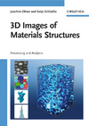
| 3D Images of Materials Structures: Processing and Analysis | |
| |
| Joachim Ohser, Katja Schladitz | |
|
|  |
 |
 |
|
|
 |
Description:
Taking and analyzing images of materials' microstructures is essential
for quality control, choice and design of all kind of products. Today,
the standard method still is to analyze 2D microscopy images. But,
insight into the 3D geometry of the microstructure of materials and
measuring its characteristics become more and more prerequisites in
order to choose and design advanced materials according to desired
product properties.
This first book on processing and analysis of 3D images of materials
structures describes how to develop and apply efficient and versatile
tools for geometric analysis and contains a detailed description of the
basics of 3d image analysis.
Table of content:
1 Lattices, adjacency of lattice points, and images
2 Image Processing
3 Image Analysis
4 Model-based image analysis
5 Modelling materials properties
Author information:
Dr. Katja Schladitz is with Fraunhofer Institute for Technomathematics
in Kaiserslautern, Germany, where she works on spatial image analysis,
modelling of microstructures and analysis of 3D images. She gives
lectures on "Image Analysis and Mathematical Morphology" and "Topology
and Image Analysis" at University of Kaiserslautern.
Professor Joachim Ohser holds a Chair at Fachhochschule Darmstadt,
Germany, and is also member of the Fraunhofer Institute for
Technomathematics in Kaiserslautern. HE also heads the working group on
quantitative texture analysis of the German Materials Society. His
research focuses on image analysis and the microstructural
characterization of metallic materials.
|
 |
| Joachim Ohser, Katja Schladitz |
| 341 Pages, November,2009 |
$180.00 now $164.95 U.S.
|
| ISBN: 978-3-527-31203-0
|
| email: info@advmedpub.net |
 |
|
|
 |
|
 |
Radiation Oncology (Clinical)
Clinical Oncology
Essential textbooks
IGRT
Neurosurgery
Prostate
Lung
Special Topics
Breast
CNS
Endocrine
Head & Neck
Gynecological
Gastrointestinal
Genitourinary
Leukemias & Lymphomas
Lung
Neuro-Oncology
Ophthalmic-Oncology
Pediatric
Prostate
Skeletal
Skin
Staging
AJCC
Color-matrix staging
Staging atlas
TNM classification
Surgical Oncology
Treatment Planning
Basic treatment planning
3-D conformal & IMRT
3-D conformal therapy
Intensity modulated RT (IMRT)
CT-simulation
Physics
Basic & clinical
Brachytherapy
General
Intra-vascular
High-dose rate
Low-dose rate
Pediatric
Quality assurance
Special procedure
Intraoperative
Stereotactic radiosurgery
Neutron capture therapy
Hyperthermia
Imaging
Abdomen
Breast
Extremities
Head & Neck
Heart
Pediatric chest
Pelvis
Spine
Thorax
CT
MRI
PET
Oncologic
Ultrasound
Oncologic Nursing
Radiobiology
Educational Materials for Residents
Physics
Radiobiology
Biological model
Clinical
Research
What's New
|


